
Artificial Intelligence-based Early-stage Glaucoma Detection with Fundus Images
Keywords
- Glaucoma, Fundus Image, Visual Field, Optical Coherence Tomography, CNN, AutoEncoder, GAN
Insights
- For patients with Glaucoma: To prevent them from blindness, they need to have an easy access to eye health examination and a reliable method to early quantize the retinal nerve fiber lesions.
- For ophthalmologists: how might we help doctors early screen out potential patients with glaucoma from fundus images
Methods
Stage 1
- Data Source: Small Public Dataset (15 fundus images from healthy patients; 15 fundus images from patients with Glaucoma)
- Data Annotations: 0 (Healthy), 1 (Glaucoma)
- Deep Learning Framework: Tensorflow (tf-slim)
- Models:
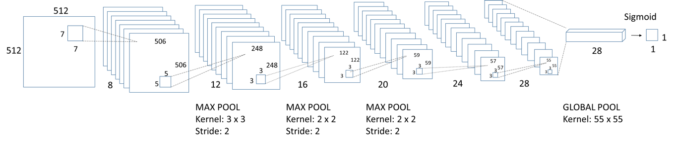
Stage 2
- Data Source: Medical Centers in Taiwan (including fundus images, visual fields, lesion map from optical coherence tomography(OCT))
- Data Annotations: Take visual fields / OCT as lesion annotations for fundus images
- Models: Inception V3 (patch-based detection method), CNN-based Auto-Encoder, VAE-GAN
Results
Stage 1
- The results are described in this conference paper
- Here are the visuals of our model
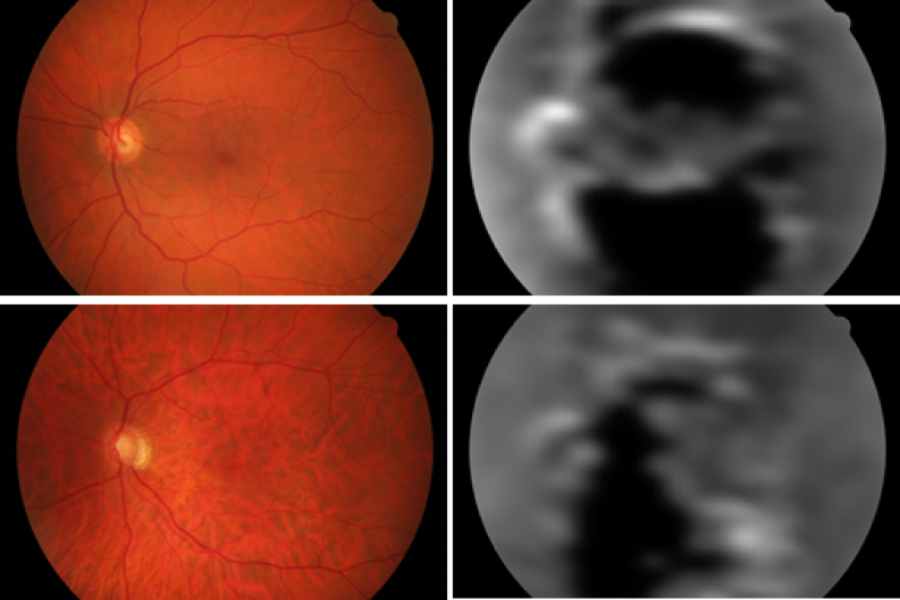
- Class Activation Map for Glaucoma
- From the ophthalmologist’s feedback, the activated regions were close to the retinal nerve fiber layer defects that they saw from the original fundus images.
Stage 2
- The experiment for using Visual Fields / OCT as the annotations for fundus images is onging. We’re using CNN-based Autoencoder and VAE-GAN to build up a model to transform fundus images into probability map of retinal nerve fiber layer defect. The results would be published in the beginning of next year.